By Dr. Alula Hailu (MD) , Diagnostic Radiology Resident
COVID19 has been the major topic for the past few months changing how we live our lives day by day. With more than 1 million confirmed cases worldwide as of today, we are learning more and more about the disease and the manifestations and different aspects of it day by day. It has shaken up the medical community and services all over the world, with radiology taking one aspect of it.
Most outpatient studies being cancelled, postponed, with shifts to more emergent imaging with revised protocol, and multiple imaging findings and diagnostic outlines being implemented at different institutions as they see fit to the local environment they are facing.
Multiple imaging findings have been documented with the COVID-19 infection on different modalities available.
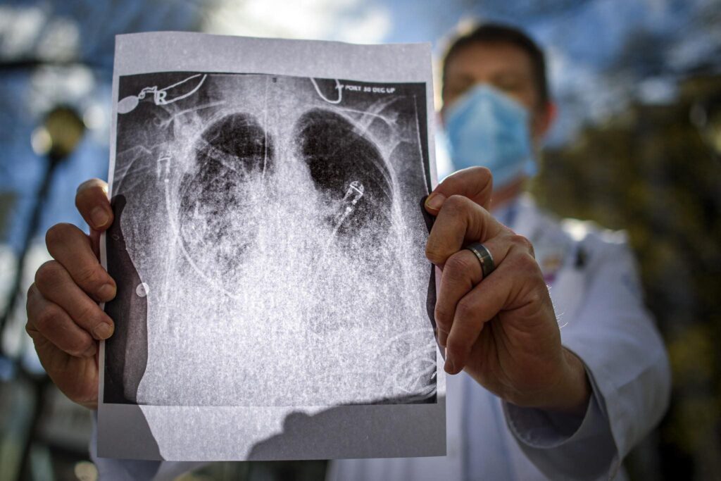
On chest radiograph, imaging findings in patients with suspected COVID-19 predominantly include bilateral infiltrates. The typical finding is patchy opacities, which tend to be predominantly peripheral and basal. The number of involved lung segments increases with more severe disease. Over time, patchy opacities may coalesce into more dense consolidation. Findings of pleural effusion, mass, cavitary lesions and lymphadenopathy are not commonly seen and might argue for an alternative or superimposed diagnosis.
Chest CT findings have been ground glass opacities (GGO), or mixed GGO and consolidation. Vascular enlargement in lesion, traction bronchiectasis, interlobular septal thickening in a crazy paving pattern. More often peripheral/subpleural, bilateral and lower lung predominant. Cavitary lesions, discrete nodules, pleural effusion and lymphadenopathy were not seen in the majority of patients who underwent CT imaging of the chest on multiple studies reported so far . Most of the CT findings are multifocal and bilateral, with fewer lesions along bronchovascular bundles. [For more robust case collection
Can Chest CT be used to Diagnose COVID-19?
Based on a study published on Feb 26, 2020 from china which included 1014 patients, 59% (601/1014) had positive RT-PCR results, and 88% (888/1014) had positive chest CT scans. The sensitivity of chest CT in suggesting COVID-19 was 97% (95%CI, 95-98%, 580/601 patients) based on positive RT-PCR results.
Other studies have shown that up to approximately 50% of patients with COVID-19 infection may have normal CT scans early during 0 to 2 days after the onset of flu-like symptoms from COVID-19. COVID-19 RT-PCR sensitivity may be as low as 60-70%; therefore patients with pneumonia due to COVID-19 may have lung abnormalities on chest CT but an initially negative RT-PCR.
However, as of now, The Centers for Disease Control (CDC) does not currently recommend CXR or CT to diagnose COVID-19. Viral testing remains the only specific method of diagnosis. Confirmation with the viral test is required, even if radiologic findings are suggestive of COVID-19 on CXR or CT.
Generally, the findings on chest imaging in COVID-19 are not specific, and overlap with other infections, including influenza, H1N1, SARS and MERS. Being in the midst of the current flu season with a much higher prevalence of influenza in the U.S. than COVID-19, further limits the specificity of CT.
Recently, there has been a report of CNS manifestations in a patient who presented with respiratory symptoms of cough and fever and altered mental status. Neurologic manifestations can occur like with any viral illness, but to a lesser extent than respiratory or gastrointestinal features reported so far. In this particular case report, the patient tested negative for influenza and positive for COVID-19 after initial evaluation. Initial imaging of the head with CT demonstrated symmetrical hypoattenuation in the medial thalami bilaterally with normal angiogram and venogram studies, at least excluding major vascular etiology to the findings.
Further imaging with MRI demonstrated peripherally enhancing lesions within the thalami bilaterally, medial temporal lobes, and sub insular regions. Based on the imaging findings, a presumptive diagnosis of Acute necrotizing encephalopathy (ANE) was made as a complication of COVID-19 infection.
ANE is a rare condition. Most cases tend to be sporadic, with a few cases of recurrent and/or familial episodes reported suggesting an inherited pattern. Patients with ANE may not have a specific symptom or classic neurological finding to suggest the diagnosis clinically. Most patients may have viral prodrome like symptoms with fever, signs of upper respiratory infection symptoms or gastroenteritis. After which patient might develop disturbance of consciousness, seizure, or focal neurological deficits. The etiology and pathogenesis are only partially clear. Usually, it develops secondary to viral infection like influenza A and B, parainfluenza, varicella and enterovirus. But a recent case report in the setting of COVID-19 pandemic has also implicated the virus SARS-COv-2 as a possible trigger. Imaging findings include thalamic, putamina, cerebral, cerebellar and brainstem areas of hypodensity, with hemorrhage and cavitation associated with worse prognosis.
“ …….This is the first reported case of COVID-19–associated acute necrotizing hemorrhagic encephalopathy. As the number of patients with COVID-19 increases worldwide, clinicians and radiologists should be watching for this presentation among patients presenting with COVID-19 and altered mental status……”3
Additional Sources:
- https://pubs.rsna.org/doi/10.1148/radiol.2020200241
- https://media.rsna.org/media/journals/specialfocus/coronavirus/images/index.html
- Poyiadji, N., Shahin, G., Noujaim, D., Stone, M., Patel, S., & Griffith, B. (2020). COVID-19–associated Acute Hemorrhagic Necrotizing Encephalopathy: CT and MRI Features. Radiology, 201187. doi: 10.1148/radiol.2020201187
- https://radiopaedia.org/articles/acute-necrotising-encephalopathy?lang=us
- https://www.rsna.org/covid-19
……………………………………………………………………………………………….
በጤና ጥበቃ የሚነገረንን መግለጫዎች በአግባቡ እንከታተል።
ባለንበት ራሳችንን እንጠብቅ፡፡
የጤና ወግ
በመረጃ የተመሰረተ ሕክምና ብቻ ።
በጥናት የተደገፈ መረጃ ብቻ እናቀርባለን።
የህክምና ባለሙያዎች ለህሙማኖቻችሁ የሚሆን የሚነበብ መረጃ ከፈለጋችሁ ይህን ግፅ ጠቁሟቸው። ህሙማኖቻችሁ እንዲያውቁት የምትፈልጉትን ነገር መረጃ ጠቅሳችሁ ፃፉልን፣ ስምችሁን ጠቅሰን እናወጣለን። የህክምና ተማሪዎች እንዲጽፉ እናበረታታለን።
በተጨማሪም በጤና ወግ ፖድካስት፣ በ ዩትዩብ ፣በ ትዊተር ፣ በ ፌስቡክ ፣ በቴሌግራም የተለያዩ የማህበራዊ ሚዲያ ገፆቻችን ታገኙናላችሁ ።


